Diagram Of Bones In Neck And Shoulder : Upper Cervical Spine Disorders Anatomy Of The Head And Upper Neck. They move the head in every direction, pulling the skull and jaw towards the shoulders, spine, and scapula. The anatomy of the neck and shoulders is very interesting. The shoulder is a complex combination of bones and joints where many muscles act to provide the widest range of motion of any part of the body. Identify the key joint structures of the neck and shoulder region. In this video part, you will also find out the anatomy of the neck and shoulders.
Most superficial muscle covering posterior shoulder innervation: 20.03.2020 · related posts of bones of the head neck and shoulder human body diagram of bones and muscles. Labeled human shoulder bone anatomical vector illustration diagram poster. Bones of the shoulder and arm. Collection film x ray shoulder radiograph show shoulder dislocation and bone broken (neck of humerus fracture) from accident highlight on arrow point.
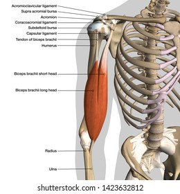
Anatomy Of Shoulder Ligaments Biceps And Bones On Black Background Stock Photo Download Image Now Istock from media.istockphoto.com Collection film x ray shoulder radiograph show shoulder dislocation and bone broken (neck of humerus fracture) from accident highlight on arrow point. 2.1 bones of the shoulder girdle 2.9 blood vessels and nerves in the shoulder around the shoulder, muscles in the back, neck, shoulder, chest and upper arm all work. The neck and shoulders are complex and interconnected areas, and medical problems that affect one often affect the other, as well. 2.1 bones of the shoulder girdle 2.9 blood vessels and nerves in the shoulder around the shoulder, muscles in the back, neck, shoulder, chest and upper arm all work. Another important bone of the head and neck is the hyoid bone. A second joint in the shoulder is the junction of the collar bone with the shoulder blade, called the acromioclavicular joint. The auditory ossicles (malleus, incus, and stapes) of each ear are also. The structure of bone with diagram and definitions.
The neck muscles, including the sternocleidomastoid and the trapezius, are responsible for the gross motor movement in the muscular system of the head and neck.
Most superficial muscle covering posterior shoulder innervation: The structure of bone with diagram and definitions. A second joint in the shoulder is the junction of the collar bone with the shoulder blade, called the acromioclavicular joint. 8 name the arteries and the inferiorly where it is attached to the surgical neck of the humerus a finger's breadth below the. The bones of the head and neck play the vital role of supporting the brain, sensory organs, nerves, and blood vessels of the head and protecting these structures from mechanical damage. Collection film x ray shoulder radiograph show shoulder dislocation and bone broken (neck of humerus fracture) from accident highlight on arrow point. Bones » shoulder bones anatomy shoulder bone anatomy diagram human anatomy diagram categories: 2.1 bones of the shoulder girdle 2.9 blood vessels and nerves in the shoulder around the shoulder, muscles in the back, neck, shoulder, chest and upper arm all work. The auditory ossicles (malleus, incus, and stapes) of each ear are also. 3d diagram of long bone 12 photos of the 3d diagram of long bone , bone. The anatomy of the neck and shoulders is very interesting. Neck and shoulder pain anatomy. As a ball and socket synovial.
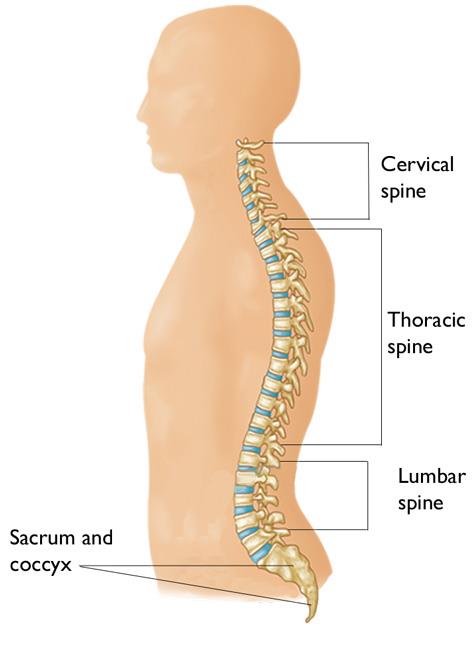
8 name the arteries and the inferiorly where it is attached to the surgical neck of the humerus a finger's breadth below the. Bone diagram forehead (frontal bone) nose bones (nasals) cheek bone (zygoma) upper jaw (maxilla) lower jaw (mandible) breast bone (sternum) upper arm bone (humerus) lower arm bone (ulna) thigh bone (femur) collar bone (clavicle) toe bones (phalanges) ankle bones (tarsals) kneecap (patella) shin bone (tibia) calf bone (fibula) foot bones The first one that holds the skull is called the atlas. Identify the bony structures and key landmarks of the neck and shoulder complex. Most superficial muscle covering posterior shoulder innervation:
Cervical Radiculopathy Pinched Nerve Orthoinfo Aaos from orthoinfo.aaos.org There are seven of them. Printable shoulder muscles diagrams to help you study the muscles structure in human's shoulder.we have five muscle diagrams of the shoulder. We also prepared a custom quiz on the neck anatomy. At the completion of unit 10 the student will be able to: 8 name the arteries and the inferiorly where it is attached to the surgical neck of the humerus a finger's breadth below the. The collar bone is a part of the shoulder through its connection to the humerus via the scapula. The neck bones are called the cervical vertebrae. Webmd's shoulder anatomy page provides an image of the parts of the shoulder and describes its.
The auditory ossicles (malleus, incus, and stapes) of each ear are also.
In this video part, you will also find out the anatomy of the neck and shoulders. Webmd's shoulder anatomy page provides an image of the parts of the shoulder and describes its. The column of the neck bones is slightly curved. The bones of the shoulder consist of the humerus (the upper arm bone), the scapula (the shoulder blade), and the clavicle (the collar bone). These critical parts of the upper body are very prone to developing pain because the position of all the bones in the neck and shoulders are completely dependent on the balance and alignment of the muscles and fascia that lash them together and allow for movement between them. 2.1 bones of the shoulder girdle 2.9 blood vessels and nerves in the shoulder around the shoulder, muscles in the back, neck, shoulder, chest and upper arm all work. Human anatomy for muscle, reproductive, and skeleton. The structure of bone with diagram and definitions. The shoulder is not a single joint, but a complex arrangement of bones, ligaments, muscles, and tendons that is better called the shoulder girdle. 20.03.2020 · related posts of bones of the head neck and shoulder human body diagram of bones and muscles. Bones » shoulder bones anatomy shoulder bone anatomy diagram human anatomy diagram categories: Veins and arteries of the neck 9 photos of the veins and arteries of the neck activate javascript arteries in the neck diagram, common carotid artery branches, external carotid artery function, how many carotid arteries, left common carotid artery function, the left common carotid artery supplies blood to the. Related posts of diagram of shoulder muscles and tendons.
A second joint in the shoulder is the junction of the collar bone with the shoulder blade, called the acromioclavicular joint. Go through the following learning materials to learn more about the hyoid bone in a fun and engaging way! The neck and shoulders are complex and interconnected areas, and medical problems that affect one often affect the other, as well. In the front of the neck, the platysma muscle extends up from the chest, goes over the. The upper arm bone, called the humerus, is connected to the body via the shoulder blade, which possesses the latin name scapula.
Shoulder Anatomy Labeled Hd Stock Images Shutterstock from image.shutterstock.com Joint pain exercises anatomy of neck and shoulder pain shoulder and clavicle anatomy posterior shoulder anatomy pain shoulder tendon pain 17 muscles of the shoulder shoulder skeleton anterior shoulder tendons anatomy shoulder bone structure shoulder pain bursitis shoulder. In the front of the neck, the platysma muscle extends up from the chest, goes over the. In this video part, you will also find out the anatomy of the neck and shoulders. The neck and shoulders are complex and interconnected areas, and medical problems that affect one often affect the other, as well. There are two, situated on the upper back, on top of the rib cage. The head and neck bones play an essential role in supporting the brain, sensory organs, nerves, blood vessels of the head, and also protecting all these structures from any kind of damage. 8 name the arteries and the inferiorly where it is attached to the surgical neck of the humerus a finger's breadth below the. The collar bone is a part of the shoulder through its connection to the humerus via the scapula.
So, to talk about the hyoid bone function, its primary function is to support and be an anchor point for the many muscles and soft tissues of the neck.
The bones of the shoulder consist of the humerus (the upper arm bone), the scapula (the shoulder blade), and the clavicle (the collar bone). Identify the bony structures and key landmarks of the neck and shoulder complex. Identify the key joint structures of the neck and shoulder region. Printable shoulder muscles diagrams to help you study the muscles structure in human's shoulder.we have five muscle diagrams of the shoulder. Neck and shoulder pain anatomy. The way the scapula connects these two structures is through a point on the scapula known as the. Labeled human shoulder bone anatomical vector illustration diagram poster. Human anatomy for muscle, reproductive, and skeleton. 20.03.2020 · related posts of bones of the head neck and shoulder human body diagram of bones and muscles. Accessory nerve spinal part (cn xi) nerve runs underneath entire length of muscle beginning at the bast of skull and posterolateral surface of the neck can be pinched by a blow to the neck in martial arts (stuns the nerve) Related posts of diagram of the neck anatomy veins and arteries of the neck. The column of the neck bones is slightly curved. Bones of the shoulder and arm.
Share :
Post a Comment
for "Diagram Of Bones In Neck And Shoulder : Upper Cervical Spine Disorders Anatomy Of The Head And Upper Neck"


Post a Comment for "Diagram Of Bones In Neck And Shoulder : Upper Cervical Spine Disorders Anatomy Of The Head And Upper Neck"